
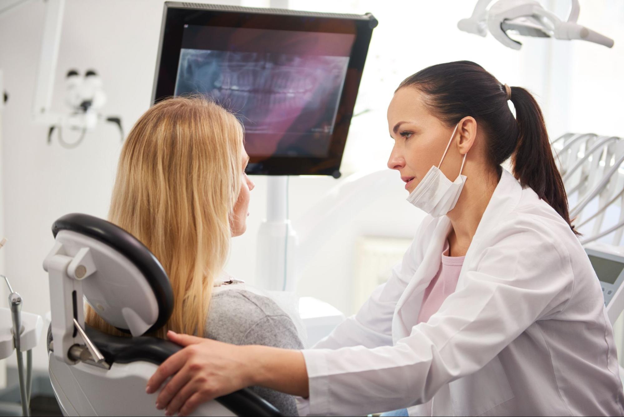
Dental radiographs, also known as X-rays, serve as an essential diagnostic tool in dentistry. They allow dentists to read and identify problems that may not be visible during a regular examination.
Dental X-rays are an essential tool that dentists use to diagnose and treat various dental issues. Incorporating dental X-rays into your oral care routine can help identify problems early on and prevent more serious complications.
So make sure to schedule regular visits with your dentist to ensure optimal oral health. So why wait for dental issues to escalate when early detection through dental X-rays can pave the way for better oral health? Visit your dentist today. Let’s dive deeper into the world of dentistry and explore how these remarkable tools enhance both prevention and treatment options for oral health problems.
Types of Dental X-Rays and Their Uses
Dental X-rays play a crucial role in detecting issues early for better oral care. There are different types of dental X-rays that dentists use to get a comprehensive view of your teeth, gums, and jaw. Let’s explore the main types and their specific uses.
Bitewing X-Rays: Detecting Cavities Between Teeth
Bitewing x-rays are one of the most commonly used types of dental x-rays. These X-rays focus on the back teeth, specifically the molars and premolars. By using bitewing X-rays, dentists can detect cavities that may be hiding between your teeth.
Cavities often develop in hard-to-reach areas where toothbrushes struggle to clean thoroughly. With bitewing X-rays, dentists can identify these hidden cavities early on, allowing for prompt treatment before they worsen. Early detection means less invasive procedures and potentially saving you from more extensive dental work down the line.
Periapical X-Rays: Examining Tooth Roots and Surrounding Bone
Periapical X-rays provide a detailed view of individual teeth from root to crown. These X-rays capture images showing the entire tooth structure, including its roots and surrounding bone. Dentists use periapical X-rays to examine specific problem areas or when they suspect an issue with a particular tooth.
By analysing periapical X-ray images, dentists can identify problems such as abscesses, cysts, impacted teeth, or any abnormalities related to tooth roots or surrounding bone structure. This type of dental X-ray helps dentists determine appropriate treatment plans tailored to address specific concerns.
Panoramic X-Rays: A Comprehensive View of Your Oral Health
Panoramic X-rays provide an overall view of your entire mouth and jaw area in a single image. This type of dental x-ray captures a broad perspective by rotating around your head. It allows dentists to assess your entire oral health, including the position and growth of teeth, jawbone density, and potential issues with your temporomandibular joint (TMJ).
With panoramic X-rays, dentists can identify problems such as impacted teeth, tumours or cysts in the jawbone, and signs of gum disease. This type of x-ray is useful for planning orthodontic treatments or evaluating the need for dental implants.
Cone Beam Computed Tomography (CBCT): Detailed 3D Imaging
Cone Beam Computed Tomography (CBCT) is a more advanced form of dental imaging that provides detailed three-dimensional images of your teeth, jawbone, nerves, and soft tissues. CBCT scans are particularly helpful when planning complex dental procedures such as dental implant placement or orthognathic surgery.
By using CBCT technology, dentists gain a comprehensive understanding of your oral anatomy. This enables them to visualise structures from multiple angles and accurately plan treatments with precision. CBCT scans are especially valuable in cases where traditional X-rays may not provide sufficient information.
Safety Considerations for Dental X-Rays
Dental x-rays play a crucial role in detecting oral issues early, enabling better oral care. However, it is natural to have concerns about radiation exposure during these procedures.
Low Levels of Radiation Exposure
Radiation exposure is often a concern. Fortunately, dental x-rays involve low levels of radiation that are considered safe for patients. The amount of radiation emitted during a dental x-ray is significantly lower compared to other medical imaging techniques like CT scans or mammograms.
Minimising Radiation Exposure
To ensure patient safety, dentists take various precautions to minimise radiation exposure during dental x-ray procedures. These measures include the use of lead aprons and thyroid collars.
Lead aprons act as protective shields, preventing unnecessary radiation from reaching the patient’s body. They are designed to cover the chest and abdominal areas, shielding vital organs from any potential harm.
Thyroid collars provide additional protection by shielding the thyroid gland located in the neck region. This collar helps reduce the risk of thyroid-related complications due to radiation exposure.
Furthermore, modern dental technology has advanced significantly over the years, allowing dentists to capture high-quality images with minimal radiation doses. Dentists now utilise digital radiography systems that require lower radiation levels compared to traditional film-based methods.
Ensuring Patient Safety
Apart from using protective gear and advanced technology, dentists follow strict guidelines and protocols to ensure patient safety during dental x-ray procedures.
Here are some additional safety measures taken:
- Proper Positioning: Dentists ensure that patients are positioned correctly for accurate imaging while minimising unnecessary exposure.
- Collimation: The use of collimators helps narrow down the X-ray beam’s size and focus on specific areas of interest only, reducing radiation scatter.
- Technique Factors: Dentists adjust the exposure settings based on individual patient needs, minimising radiation while still obtaining clear diagnostic images.
- Limiting Frequency: Dentists only recommend dental X-rays when necessary, considering factors such as a patient’s oral health history and specific concerns.
By implementing these safety measures, dentists prioritise patient well-being and minimise any potential risks associated with dental x-rays.
Importance of Early Detection in Oral Health
Early detection plays a crucial role in maintaining good oral health. By identifying potential issues at their earliest stages, individuals can receive timely treatment, preventing further damage or complications. Regular dental check-ups that include dental X-rays enable dentists to catch problems before they become severe.
Detecting oral health issues early can save patients from pain and discomfort in the long run. Tooth decay, gum disease, and even cancer are among the common oral health problems that can be detected early through regular dental examinations. Dental X-rays provide valuable insights into the underlying structures of teeth and gums, helping dentists identify signs of diseases such as bone loss or infections.
Tooth decay is a prevalent issue that affects people of all ages. It occurs when bacteria in the mouth produce acids that erode tooth enamel over time. With early detection through dental X-rays, dentists can identify areas of decay before they develop into cavities. Prompt treatment at this stage may involve simple interventions like fluoride treatment or dental fillings, preventing the need for more extensive procedures like root canals or extractions.
Gum disease is another condition that benefits greatly from early detection. Often caused by poor oral hygiene habits and plaque buildup, gum disease can lead to serious complications if left untreated. Dental x-rays help detect signs of gum disease such as bone loss around the teeth or deep periodontal pockets where bacteria thrive. Early intervention with professional cleanings and improved oral hygiene practices can halt the progression of gum disease and prevent tooth loss.
Cancer screening is an essential component of routine dental exams that includes dental x-rays. These screenings allow dentists to identify any suspicious lesions or abnormalities in the mouth that may indicate oral cancer. Detecting cancer early significantly improves treatment outcomes and increases survival rates.
In addition to detecting specific conditions, dental X-rays also provide valuable information about overall oral health. They reveal hidden issues beneath the surface that may not be visible during a regular examination. Dentists can identify impacted teeth, cysts, or abnormalities in the jawbone that may require further evaluation or treatment.
How Dental X-Rays Help Detect Oral Health Issues
Dental X-rays, also known as radiographs, play a vital role in identifying and diagnosing dental problems that may not be visible to the naked eye. By capturing detailed images of the teeth and surrounding structures, these diagnostic tools enable dentists to detect issues early on and develop effective treatment plans. Let’s explore how dental X-rays assist in uncovering hidden dental issues and contribute to better oral care.
Revealing Hidden Problems
One of the primary benefits of dental X-rays is their ability to reveal hidden dental problems. While routine examinations can identify some issues, certain conditions may remain undetected without the help of radiographs. Dental X-rays expose tooth decay that may be lurking beneath the surface, allowing dentists to address it promptly before it worsens. Moreover, they can unveil signs of gum disease, infections, or impacted teeth that might otherwise go unnoticed.
Accurate Diagnosis with Detailed Images
The detailed images provided by dental X-rays are invaluable for accurate diagnosis. These images allow dentists to examine teeth from various angles and assess their condition thoroughly. By closely analysing the radiographs, dentists can identify cavities at their earliest stages when they are smaller and easier to treat. Dental X-rays aid in evaluating bone density and detecting any abnormalities or irregularities that could indicate underlying problems.
Developing Effective Treatment Plans
Detecting dental issues beneath the surface is crucial for developing effective treatment plans. Dental X-rays provide essential information about the extent of a problem and its impact on adjacent teeth or supporting structures. Armed with this knowledge, dentists can formulate appropriate treatment strategies tailored to each patient’s needs. For instance, if an impacted tooth is identified through a radiograph, a dentist can plan for its extraction before it causes further complications.
Benefits of Digital X-Rays over Traditional X-Rays
Digital X-ray technology has revolutionised the field of dentistry, offering numerous benefits over traditional film-based systems. With advancements in digital imaging, dental professionals can now detect issues early on for better oral care. Let’s explore the upsides of digital X-rays and why they are preferred in modern dental practices.
Reduced Radiation Exposure
One significant advantage that digital x-ray technology brings is a substantial reduction in radiation exposure compared to traditional methods. Studies have shown that digital X-rays can lower radiation levels by up to 80%. This decrease in radiation ensures patient safety without compromising the accuracy of diagnostic results.
Instant Analysis and Diagnosis
Gone are the days when patients had to wait for their x-ray films to be developed. With digital imaging, dentists can view images instantly on a computer screen, enabling immediate analysis and diagnosis. This real-time access allows for prompt treatment planning and enhances patient communication as dentists can explain their findings while examining the images together.
Improved Efficiency with Electronic Storage
The advent of digital X-rays has eliminated the need for physical storage space required by traditional film-based systems. Digital images can be easily stored electronically, saving valuable office space and reducing clutter. Moreover, retrieving specific images becomes effortless with just a few clicks, streamlining workflow efficiency within dental practices.
Enhanced Image Sharing Capabilities
Sharing diagnostic information between dental professionals is crucial for effective collaboration and referral purposes. Digital X-rays facilitate seamless sharing through electronic means such as email or secure file transfer protocols. This efficient exchange of information expedites consultations between specialists, leading to more comprehensive treatment plans for patients.
In addition to these primary benefits, there are other advantages associated with digital x-ray technology worth mentioning:
- Enhanced Image Quality: Digital imaging provides superior image quality compared to traditional methods, allowing dentists to detect even minor abnormalities accurately.
- Advanced Imaging Techniques: Digital X-rays enable the use of tomography, a technique that provides three-dimensional images of oral structures. This advanced imaging aids in precise diagnoses and treatment planning.
- Environmentally Friendly: Unlike traditional film-based systems that require chemicals for development, digital X-rays are more environmentally friendly as they eliminate the need for toxic processing solutions.
Precautions Taken by Dentists for Safe Dental X-Rays
Dentists prioritise patient safety and follow strict guidelines to ensure that dental x-ray procedures are conducted safely. By implementing proper positioning of the x-ray equipment and taking protective measures, dentists minimise radiation exposure risks. Regular maintenance and calibration of X-ray machines are crucial to guarantee accurate results and safe operation.
Patient safety is paramount. Dentists take several precautions to protect patients from unnecessary radiation exposure. One essential precaution is ensuring that the X-ray equipment is positioned correctly. By carefully aligning the machine, dentists can target specific areas of the mouth while minimising exposure to other parts of the body.
To further enhance safety, dentists employ protective measures during dental x-ray procedures. Patients are provided with lead aprons or thyroid collars to shield sensitive areas from radiation. These protective garments act as a barrier, reducing the absorption of harmful rays by vital organs such as the thyroid gland.
Regular maintenance and calibration of X-ray machines play a vital role in ensuring both accuracy and safety during dental X-rays. Dentists adhere to strict schedules for servicing their equipment, which includes inspecting components for any signs of wear or damage. This proactive approach helps identify potential issues before they compromise patient safety or affect the quality of images produced.
Calibration is another critical aspect that dentists consider when conducting dental x-rays. Properly calibrated machines provide accurate readings, enabling dentists to detect oral health issues early on with precision. Calibration involves adjusting various settings within the machine to optimise image quality while minimising radiation output.
In addition to these precautions, dentists also stay up-to-date with advancements in technology and best practices related to dental radiography. They undergo continuous education and training programs focused on radiation safety protocols and techniques for minimising exposure risks.
By following these precautions diligently, dentists ensure that patients receive the benefits of dental X-rays without compromising their overall well-being. Early detection of oral health issues through X-rays allows dentists to provide timely and effective treatments, leading to better oral care outcomes.
The Significance of Dental X-Rays for Optimal Oral Care
In conclusion, dental X-rays play a crucial role in ensuring optimal oral care. By detecting issues early on, these diagnostic tools enable dentists to identify problems that may not be visible to the naked eye. With various types of dental X-rays available, dentists can target specific areas and gain valuable insights into your oral health. This early detection is essential because it allows for timely treatment and prevents further complications down the road.
To ensure you receive the best possible oral care, make sure to schedule regular dental check-ups that include X-rays. These visits will help your dentist detect any potential issues before they worsen, saving you from unnecessary pain and expenses in the future. Remember, prevention is always better than cure when it comes to your oral health!
FAQ
Can dental x-rays detect cavities?
Yes, dental x-rays are an effective tool for detecting cavities. They can reveal cavities between teeth or beneath fillings that may not be visible during a visual examination.
Are dental x-rays safe?
Yes, dental x-rays are considered safe with minimal radiation exposure. Dentists take precautions such as using lead aprons and collars to protect patients from unnecessary radiation.
How often should I get dental x-rays?
The frequency of dental X-rays depends on your individual oral health needs. Generally, adults may require them every 1-2 years, while children or those at higher risk may need them more frequently.
Do digital X-rays have any advantages over traditional X-rays?
Yes, digital X-rays offer several advantages over traditional film-based ones. They provide instant results, emit less radiation, and allow for easier storage and sharing of images.
Can pregnant women have dental x-rays?
Pregnant women should avoid dental x-rays unless absolutely necessary. However, if there is an urgent need for an x-ray due to a dental emergency, appropriate precautions can be taken to minimise radiation exposure. It is important to inform your dentist if you are pregnant.